

Yes – a pelvic ultrasound can detect pregnancy by showing the baby’s early development inside the womb. In early pregnancy the scan will reveal the gestational sac (the fluid-filled sac where the embryo grows), the yolk sac (a small structure that feeds the embryo), and eventually the fetal pole (the embryo itself) and heartbeat. In fact, medical guidelines say an ultrasound can see a gestational sac as early as about 4½–5 weeks of gestation. By roughly 6 weeks, most pregnancies show a fetal pole and heartbeat on a good scan. Pelvic ultrasound is therefore routinely used in the first trimester to confirm a pregnancy (and that it is inside the uterus), to estimate how far along you are, and to check that everything looks healthy.
How a Pelvic Ultrasound Works
A pelvic ultrasound is a safe, painless scan that uses high-frequency sound waves (like sonar) to create images of the uterus, ovaries, and developing baby. It works by moving a handheld probe (transducer) over the lower belly or inside the vagina; the probe sends sound waves into the body, and a computer turns the echoes into real-time pictures. Obstetric ultrasound uses no radiation and has no known harmful effects, making it the preferred method to check on early pregnancy.
- Transabdominal ultrasound: This is the “belly” scan. The patient usually lies down with a full bladder, and the probe glides over the abdomen. It’s simple and painless, but in very early pregnancy it may not be sensitive enough to see tiny structures.
- Transvaginal ultrasound: In the first trimester, doctors often prefer this approach for clearer images. A slim probe is gently inserted into the vagina, bringing it closer to the uterus. This higher-frequency scan gives a more magnified view of the pregnancy. (For example, one medical review notes that transvaginal ultrasound is “the modality of choice” for confirming a new pregnancy because it can see small details better.) No full bladder is needed for a transvaginal scan, and it can often pick up a pregnancy about a week or two earlier than an abdominal scan.
Because pelvic ultrasound is noninvasive and uses sound, you won’t feel pain (aside from mild pressure from the probe). A clear gel is applied so sound waves travel easily, and the images (sometimes fuzzy 2D grey-scale pictures) show up on a monitor. You can usually see the gestational sac as a small black circle in the uterine lining once it’s about 5 weeks old.
Early Pregnancy Signs on Ultrasound
Pelvic ultrasound is very effective at early detection of pregnancy. Here’s roughly what it can show in the first few weeks:
- Gestational sac: Usually the first thing you see. At about 4½–5 weeks, a tiny round fluid-filled sac (gestational sac) becomes visible inside the uterus. This is the hallmark of an intrauterine pregnancy.
- Yolk sac: By roughly 5–5.5 weeks the yolk sac appears as a small circle inside the gestational sac. It feeds the embryo in the very early days.
- Fetal pole (embryo): Around 5½–6 weeks you’ll typically see a tiny embryo (often just a millimeter or two long) adjacent to the yolk sac. As it grows, doctors measure its crown-to-rump length to estimate gestational age.
- Heartbeat: Once the fetal pole is visible, you can usually detect a heartbeat soon after – often around 6 weeks or a bit later. Seeing the fetal heartbeat is a clear sign of a viable pregnancy.
- Other features: By the end of the first trimester, a routine pelvic ultrasound can show the developing baby’s limbs, spine, and placenta. Even early on, the ultrasound can reveal multiple sacs if you’re carrying twins or more.
In short, pelvic ultrasound lets your doctor see the baby grow. It can pick up even very early signs of life in the uterus and then follow those signs as the pregnancy progresses. Medical sources confirm these milestones: for example, one review notes that by 6 weeks the embryo and cardiac pulsations are usually visible.
Why Doctors Use Pelvic Ultrasound
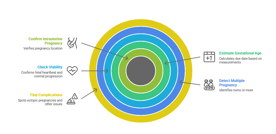
A pelvic ultrasound isn’t just about confirming you’re pregnant; it answers important health questions too. Here are some key reasons an early-pregnancy scan is done:
- Confirm intrauterine pregnancy: It verifies that the pregnancy is in the uterus (not an ectopic in the tube). Spotting the gestational sac and later the fetus in the uterus is the surest way to confirm pregnancy location.
- Estimate how far along you are: By measuring the gestational sac, yolk sac, and embryo size, the sonographer can estimate gestational age. This helps calculate a more accurate due date if you’re unsure of your last period. (The crown–rump length at about 6–8 weeks is one of the most accurate ways to date a pregnancy.)
- Check viability: Hearing or seeing the fetal heartbeat on ultrasound reassures both patient and doctor that the pregnancy is progressing normally. It also rules out miscarriage in cases of bleeding or cramping.
- Detect multiple pregnancy: If you’re having twins or more, ultrasound will show multiple sacs or embryos at the start. This allows early monitoring and care.
- Find complications: An ultrasound can spot conditions like an ectopic pregnancy (pregnancy outside the uterus, usually a medical emergency) or a blighted ovum (empty sac without an embryo). It can also identify ovarian cysts or uterine issues that might affect the pregnancy.
- General health check: The scan looks at the mother’s uterus and ovaries as well – checking for fibroids, cysts, or other conditions that might require attention.
Radiology experts agree that ultrasound is ideally suited for these tasks. For instance, one authoritative source lists “establishing the presence of a living embryo/fetus,” estimating the age of the pregnancy, and diagnosing conditions like ectopic pregnancy as common uses of obstetric ultrasound. In practice, if a patient has a positive pregnancy test, doctors often follow up quickly with a pelvic ultrasound to confirm everything looks normal.
Safety, Preparation, and What to Expect
Pelvic ultrasound is a routine, low-risk procedure. It uses sound waves (no X-rays), so it poses no radiation risk to you or the baby. You may be asked to drink water and come with a full bladder if you’re having an abdominal scan – the full bladder helps push the uterus into view. For a transvaginal scan, you’ll empty your bladder and lie comfortably with the probe inserted (covered with a disposable sheath) for better images.
Typically, the exam takes only a few minutes. You might feel slight pressure when the probe is moved or when the vaginal probe is inserted, but it shouldn’t hurt. The gel on your belly may feel cold at first. You’ll be able to see the black-and-white images on the screen, and the sonographer or doctor will point out the gestational sac, fetal pole, or heartbeat. It can be an emotional moment for many expectant parents!
If the scan is too early in pregnancy and nothing is visible yet, the doctor may ask to repeat it in 1–2 weeks. This sometimes happens if the test was done right at 4 or 5 weeks. In general, doctors recommend the first ultrasound around 6–8 weeks of pregnancy, when the baby’s development is more readily visible. (In fact, one obstetrics group notes that ultrasounds can be performed as early as 5 weeks, but waiting until 6–8 weeks often gives a clearer picture.)
Conclusion
In summary, a pelvic ultrasound is a powerful tool for detecting and monitoring early pregnancy. It can confirm pregnancy by visualizing the gestational sac and embryo, often as early as 5 weeks. By the end of the first trimester it will clearly show the developing baby’s heartbeat and anatomy. This scan is safe, painless, and routinely used by obstetricians to make sure both mother and baby are healthy and on track. If you have a positive pregnancy test or pregnancy symptoms and wonder about scanning, rest assured that modern pelvic ultrasound is the standard method to check early pregnancy status and guide your care.