

Pregnancy ultrasound reports can seem full of medical jargon, but you can get the main points by focusing on the Findings and Impression/Conclusion sections. These sections summarize what the technician saw and conclude whether everything looks normal. Key numbers to check include the baby’s measurements – for example, Crown–Rump Length (CRL), which is the baby’s length from head to bottom (used to date early pregnancy) – and Gestational Age (GA), usually shown in weeks. The report will often list an Estimated Due Date (EDD) based on these measurements. It may also note placenta and amniotic fluid details. In the ultrasound image itself, remember that black areas are fluid, gray is soft tissue, and white is bone. Reading the report with these points in mind will give you a clear overview of your baby’s growth.
Key Report Sections: Findings and Impression
The Findings section is the detailed part of the report. It lists what was seen – organ sizes, fluid pockets, and whether each looks normal or not. The Impression (or Conclusion/Summary) is a short summary of those findings. Doctors often say things like “normal” or “unremarkable” here if everything looks okay. For expectant parents, the Impression is especially important because it highlights the big points in plain terms. For example, it usually mentions the baby’s heartbeat, how many weeks along you are (GA), whether it’s a single baby or multiples, and any notes on the placenta or fluid. In short: if the Impression says your pregnancy is “normal” or “within normal limits,” that generally means no concerns were found.
Important Measurements and Terms
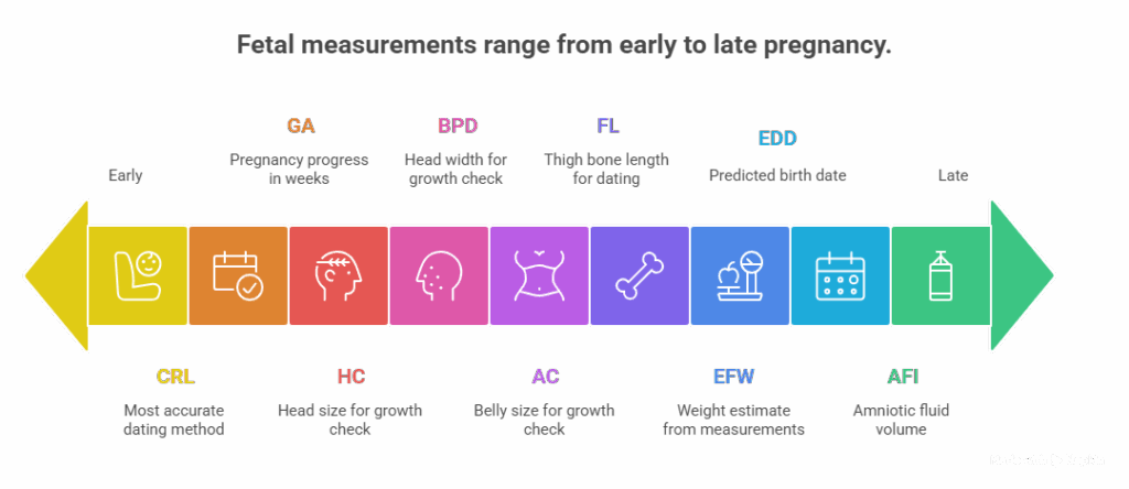
Ultrasound reports use common acronyms for fetal measurements. Here are some you’ll see often (with their meanings):
- CRL (Crown–Rump Length): Baby’s length from top of head to bottom. It is the most accurate way to date a pregnancy in the first trimester (about 6–13 weeks).
- GA (Gestational Age): How far along the pregnancy is, in weeks and days. This is usually based on your last menstrual period and/or the measurements seen on ultrasound.
- HC (Head Circumference): The size around the baby’s head. Used (with other measurements) to check growth. (Usually measured after the first trimester).
- AC (Abdominal Circumference): The measurement around the baby’s belly. It helps assess growth and size.
- FL (Femur Length): The length of the baby’s thigh bone. Used to estimate gestational age later in pregnancy.
- BPD (Biparietal Diameter): The width of the baby’s head (distance between the two sides). This is another head size measure, checked from mid-pregnancy onward.
- EFW (Estimated Fetal Weight): An estimate of the baby’s weight, calculated from HC, AC, FL and other measurements.
- EDD (Estimated Due Date): The predicted birth date, calculated from LMP and/or ultrasound measurements.
- AFI or AF (Amniotic Fluid Index): How much amniotic fluid is around the baby. The report will note if fluid is at a normal level or too low/high.
- Placenta Location: Where the placenta is attached (e.g. “anterior” = front, “posterior” = back, or “low-lying”). A normal report will note something like “posterior placenta, normal in appearance”. If it says “placenta previa” or “low-lying placenta,” that means the placenta is near the cervix, which your doctor will watch.
- Fetal Heart Rate (FHR): The baby’s heartbeat, usually given in beats per minute. A normal rate in early pregnancy is often around 120–160 bpm.
Other terms you might see on an early scan report include the Gestational Sac (GS) – the fluid-filled sac around the embryo – and the Yolk Sac (YS), an early source of nutrients. The report may also say singleton (one baby) or multiple pregnancy (twins, etc). If the report uses any term you don’t recognize (like “Nuchal Translucency,” “chorion,” etc.), make a note to ask your doctor to explain it.
Interpreting the Ultrasound Image
Although your doctor will describe the findings, it’s also helpful to know what the scan image shows. An ultrasound picture is a black-and-white (“grayscale”) image: black areas are fluid (amniotic fluid or the bladder), gray areas are soft tissues (your baby’s organs and muscles), and white areas are very dense structures like bone. For example, in a typical prenatal scan the top part of the image (closest to the probe on your belly) often looks gray because it’s showing the uterine wall and tissue, and beneath that you’ll usually see a black region of amniotic fluid.
You may also see color on the screen – that’s Doppler ultrasound, which shows blood flow. Don’t worry if you see red and blue streaks: red usually means blood moving toward the probe, and blue means blood moving away. These colors are just tools the technician uses to check blood flow in the placenta, umbilical cord, or baby’s heart; they don’t mean anything bad by themselves. The main thing to remember is the black/gray/white rule for fluid, tissue, and bone. (If you’re curious, images are often mirror-like, so the baby’s left is usually on the left side of the image.)
What You’ll See at Different Stages
Early pregnancy (5–12 weeks) – The report will confirm you have an intrauterine pregnancy. It will mention the gestational sac in the uterus (a dark circle) and the fetal pole (tiny embryo) with a flickering heartbeat. It will give a CRL measurement – for example, a CRL around 1.2 cm usually means ~7 weeks gestation. A normal report at this stage might say something like “viable intrauterine pregnancy of 7 weeks, fetal heart rate 150 bpm.” The yolk sac might also be noted. If everything is on track, the Impression might simply read “single live intrauterine pregnancy” with the corresponding number of weeks.
Second trimester (13–26 weeks) – The scan becomes more detailed. You’ll start seeing your baby’s anatomy: head, body, spine, arms and legs. The report will list growth measurements such as BPD, HC, AC, and FL (head width, head circumference, belly circumference, femur length). The technician checks these against normal charts. For example, [24] notes that in the second trimester “BPD” (head width), “AC” (abdominal circumference), and “FL” (thigh length) are important to track growth. The report will also describe major organs (heart, kidneys, brain) as normal or flag if something is unusual. You may even learn baby’s sex if you want. By the end of this scan, you’ll often get an updated EDD (if it changed) and a clear statement of fetal growth.
Third trimester (27+ weeks) – The baby is bigger and almost fully formed. The focus is on growth and position. The report will still show head, belly, and limb measurements, and often an EFW (estimated fetal weight) is given to estimate how much the baby weighs now. The Amniotic Fluid Index (AFI) or fluid level will be noted – too little or too much fluid can be a concern. [24] points out that in the third trimester scans, estimated weight and amniotic fluid volume are key terms. The placenta and umbilical cord are checked (for example, the report might say “anterior placenta, normal appearance”). The impression might focus on baby’s position (head down or not) and whether growth is on track.
Common Ultrasound Image Clues
If you have a photo or live image from your ultrasound, here are some quick tips:
- Find the baby: The baby will be the gray/white blob inside a black fluid space. In early scans, it looks like a little bean shape. In later scans, you’ll see limbs, spine, etc..
- Color on the screen: Red/blue areas mean blood flow (to/from the probe).
- Numbers on the screen: Sometimes the machine prints small labels or numbers around the picture. Those are usually camera settings, date/time, or basic measurements. Don’t worry if you don’t recognize them – your report will explain any important numbers.
Key Takeaways
- Impression is key: Start by reading the Impression/Conclusion. If it says “normal” or “no abnormalities,” that generally means everything in the findings looks good.
- Focus on major items: Make sure a heartbeat is noted (“cardiac activity present”) and that the gestational age (GA) matches how far along you think you are. Check how many babies were found (single or multiples), and note the estimated due date (EDD).
- Use the report for conversation: Don’t fret over unfamiliar terms. For example, a “normal” report includes seeing a gestational sac, a fetus with a heartbeat, and size measurements that match the GA. If your report is normal, it will list those reassuring findings. If there are any concerns, the report should explicitly say what they are.
- Ask questions: Write down anything confusing and discuss it with your doctor or midwife. They can explain terms like “placenta previa” or “oligohydramnios” (low fluid) in plain language. Remember, ultrasound reports are for doctors, but your healthcare team wants you to understand your baby’s progress.
Pregnancy ultrasounds are a routine, safe way to check on your baby (they use sound waves, not X-rays). By knowing what the key measurements and sections mean, you can read the report with confidence. Keep in mind that each pregnancy is unique – the report is a tool to see how yours is growing. Use it as a guide, and always feel free to ask your care provider for clarity if you’re unsure about anything.